POSITRON EMISSION TOMOGRAPHY
PET is minimally invasive and quantitative imaging modality which uses molecules labelled with short-lived positron-emitting radioisotopes to visualize and measure rates of biochemical processes (e.g. cellular metabolism, cell proliferation, enzyme reactions, ligand-receptor interactions) in living animals and humans. PET is routinely integrated with CT and MRI in hybrid PET/CT and PET/MRI systems. The specialty of the Finnish Biomedical Imaging Node in PET imaging is exceptionally broad PET tracer collection for both human and animal studies.
The Finnish Biomedical Imaging Node produces PET tracers onsite and offers exceptionally broad collection of different PET tracers and applications for both animal and human studies. The most commonly used tracers in different research fields are listed below, but as our PET tracer portfolio is constantly being streamlined with new tracers developed in-house, don’t hesitate to ask for other options – there are many!
CARDIOVASCULAR AND METABOLIC PET IMAGING
The cardiovascular PET research in the Finnish Biomedical Imaging Node is focused on diagnostics, characterization, and monitoring coronary artery disease, heart failure, and inflammatory processes of the cardiovascular system. The Node’s metabolic PET applications provide various opportunities for studying insulin resistance and pancreatic beta-cell dysfunction in obesity, metabolic diseases, and T2 diabetes. The Node is also specialized in investigating the role of brown adipose tissue in human metabolism and the effects of physical activity/inactivity on tissue specific and whole-body metabolism. Animal models for cardiovascular and metabolic diseases ranging from mice to pigs are routinely available.
PET TRACERS FOR CARDIOVASCULAR AND METABOLIC IMAGING (SELECTED EXAMPLES)
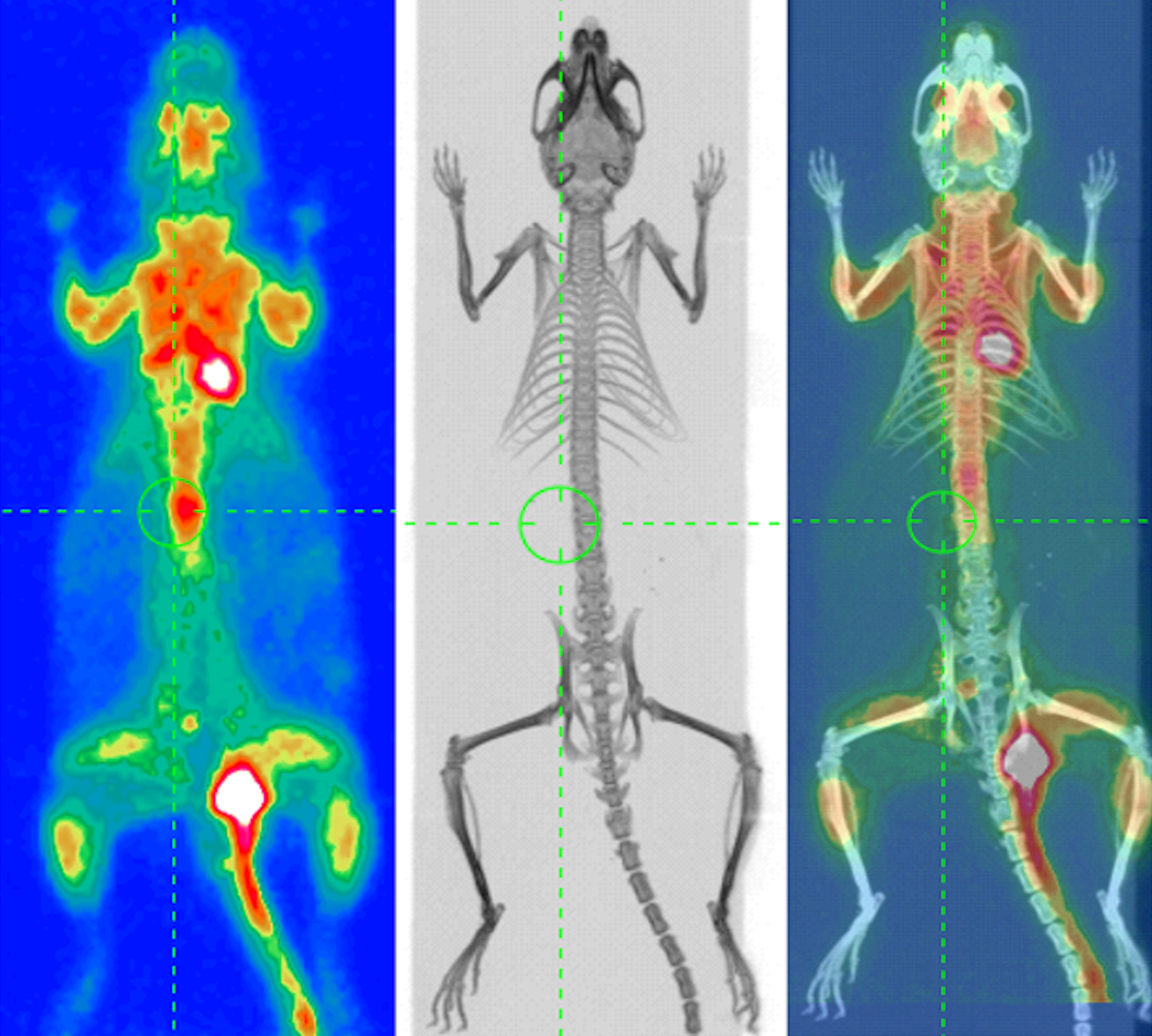
- [18F]FDG ⎯ Glucose metabolism
- [15O]Water ⎯ Blood flow, perfusion
- [15O]O2 ⎯ Oxygen consumption
- [18F]FTHA ⎯ Fatty acid metabolism
- [11C]Acetate ⎯ Oxidative metabolism
- [18F]EF5 ⎯ Hypoxia
- [68Ga]-DOTA-Siglec-9 ⎯ Leukocyte trafficking (inflammation)
BRAIN PET IMAGING
The brain PET applications in the Finnish Biomedical Imaging Node are focused on neurophysiology of aging brain and pathophysiology of neurogenerative diseases, as well as neuroreceptor systems in health and disease. The Node also provides applications for investigation of molecular, structural, and functional processes (e.g. neuroinflammation) of different psychiatric and neurological disorders, as well as the neurobiology of human behavior, emotions, and cognitive functions.
PET TRACERS FOR BRAIN IMAGING (SELECTED EXAMPLES)
- [11C](R)-PK11105 ⎯ Microglial activity (neuroinflammation)
- [11C]PIB ⎯ Amyloidosis (β amyloid plaque)
- [11C]Raclopride ⎯ Dopamine D2 receptors
- [11C]PBR28 ⎯ Microglial activity (neuroinflammation)
- [11C]Carfentanil ⎯ µ opioid receptors
- [18F]FDOPA ⎯ Dopaminergic function
- [11C]UCB-J ⎯ Synaptic density
ONCOLOGICAL PET IMAGING
The oncological PET expertise of the Finnish Biomedical Imaging Node offers opportunities to assess and validate new hybrid imaging methods, PET tracers, and image acquisition protocols in cancer diagnostics. The Node is also specialized in developing new theranostic approaches for various types of cancers, such as prostate and breast cancer, neuroendocrine and other somatostatin receptor positive tumors, and malignant lymphoma.
PET TRACERS FOR CANCER IMAGING (SELECTED EXAMPLES)
- [18F]FDG ⎯ Glucose metabolism
- [11C]Choline ⎯ Phospholipid metabolism, cell proliferation
- [11C]Acetate ⎯ Fatty acid metabolism
- [11C]Methionine ⎯ Amino acid transport
- [11C]Metomidate ⎯ 11β-hydroxylase activity
- [18F]EF5 ⎯ Hypoxia
- [18F]DOPA ⎯ Dopaminergic function
- [68Ga]RGD – Angiogenesis
- [68Ga]DOTANOC – Somatostatin receptors
- [18F]PSMA ⎯ Prostate-specific membrane antigen
The Finnish Biomedical Imaging Node is fully equipped for synthesis and development of various PET radiotracers. The Node houses several cyclotrons and generators for radionuclide production (e.g. 15O, 18F, 11C, 64Cu, 68Ga), GMP compliant radiochemistry facilities with automated synthesis devices, and expertise to develop PET tracers from chemistry through in vitro and preclinical tests to clinical use. Several new PET tracers are tested annually, and many of them are being developed in collaboration with major pharmaceutical companies. Therefore, the Finnish Biomedical Imaging Node offers perfect environment to explore and optimize new potential radioligands which are continuously needed in research and the clinic.
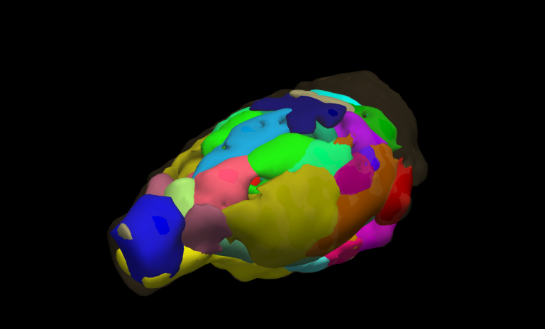
PRECLINICAL HIGH-FIELD MRI
High-field MRI uses radiofrequency waves and a strong magnetic field to provide anatomical images of internal organs and tissues as well as information of dynamic, functional and metabolic processes in vivo. Specialized techniques allow examination of brain function and cardiac function, fat distribution, and brain microstructure and metabolism. Finnish Biomedical Imaging Node is specialized in functional MRI methods in awake animals and hyperpolarized MRI for metabolic imaging.
The Finnish Biomedical Imaging Node provides unique fMRI technology to study intrinsic and spontaneous brain activities in awake animals both in rest and in response to stimuli. The technique allows for example diverse genetic, electrical, and pharmacologic manipulations and at the same time overcomes the interfering effects of anesthetics. The unique technique is minimally stressful to animals and tolerant to body movement related image artefacts. It can also be combined with various brain stimulation or electrophysiological recordings providing flexible study designs.
The Finnish Biomedical Imaging Node houses DNP-hyperpolarizer for 13C-labeled probes, such as pyruvate. Hyperpolarization increases MR sensitivity of 13C nuclei over 10000-fold for 1-2 minutes which allows real-time imaging of pathway-specific metabolic processes, where the injected probe is observed separately from its metabolites. Therefore, hyperpolarized MRI has applications in many diseases with altered metabolism including cancer, cardiovascular disease, diabetes, and a variety of inflammatory conditions.
MAGNETOENCEPHALOGRAPHY (MEG)
MEG is a functional brain imaging technology that measures noninvasively the weak magnetic fields produced by the electrical activity of neurons. MEG is optimally suited for detecting neuronal dynamics in the cerebral cortex, which has made it a powerful tool in basic neuroscience research, with well-established clinical applications in epilepsy and brain surgery. Currently, the Finnish Biomedical Imaging Node is the only Euro-BioImaging Node offering MEG.
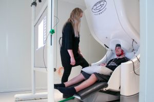
The Finnish Biomedical Imaging Node provides MEG-optimized experimental designs coupled with a wide range of sensory, motor and cognitive stimulus systems. The offered protocols can be used to study for example sensorimotor and proprioceptive brain activity related to body movements, cortical networks in language processing, and neuronal signaling of cognitive functions in health and disease. MEG is routinely combined with other complementary measures of human neurophysiology such as fMRI, EEG, TMS, and eye-tracking as well as with diverse behavioral measures.
The Finnish Biomedical Imaging Node provide opportunities for MEG related imaging and neurotechnological development. New types of MEG sensors, hybrid MEG-MR systems, and next-generation MEG stimulator systems and monitoring devices are actively being developed in house.
OPTICAL INTRAVITAL IMAGING
Optical intravital microscopy enables imaging of living animals down to subcellular, submicrometer resolution. It allows examination of various cellular processes, such as cell behavior, how cells interact with other cells, and how cells respond to various stimuli in their microenvironment both in anesthetized and awake animals. The most widely used optical intravital microscopy set-ups for preclinical imaging are based on multiphoton microscopy. Finnish Biomedical Imaging Node offers wide variety of intravital imaging set-ups for both anesthetized and awake animals.
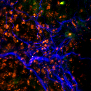
The Finnish Biomedical Imaging Node provides multiphoton, widefield and intrinsic signal optical intravital imaging set-ups for studying e.g. ear skin vasculature, paw skin, brain, lymph node/mammary gland, tumors, and externalized internal organs such as the intestine, liver and spleen, together with protocols and adapters for both anesthetized and behaving animals (brain imaging). These techniques can be used to study for example vascular networks, blood flow and leakage, cell tracking, and morphological changes in longitudinal settings.
Advanced neuronal set-ups for brain imaging allow for example dynamic imaging of calcium- or voltage-sensitive proteins, micro-anatomic imaging of dendritic spines and mitochondria, and blood oxygenation and blood flow rate imaging. Brain imaging can also be performed in freely moving animals by using in-house developed home-cage device, where the animal can be exposed to cues, perform tasks or interact with other animals and coupled with physiological recordings.