Skip to content
Finnish Advanced Microscopy Node
  • Home
  • About Us
    • FiAM Node
    • Impact and viability
    • Testimonials
    • FiAM logos
  • Services
    • Technologies
    • Highlighted Technologies
    • Additional Services
  • Access to services
    • How to access services
    • User Fees
    • Funding for user access
  • Publications
  • Links
    • National partners
    • International partners
    • Resources
    • Software
  • Contact us
    • Node Partners
    • General Inquiries

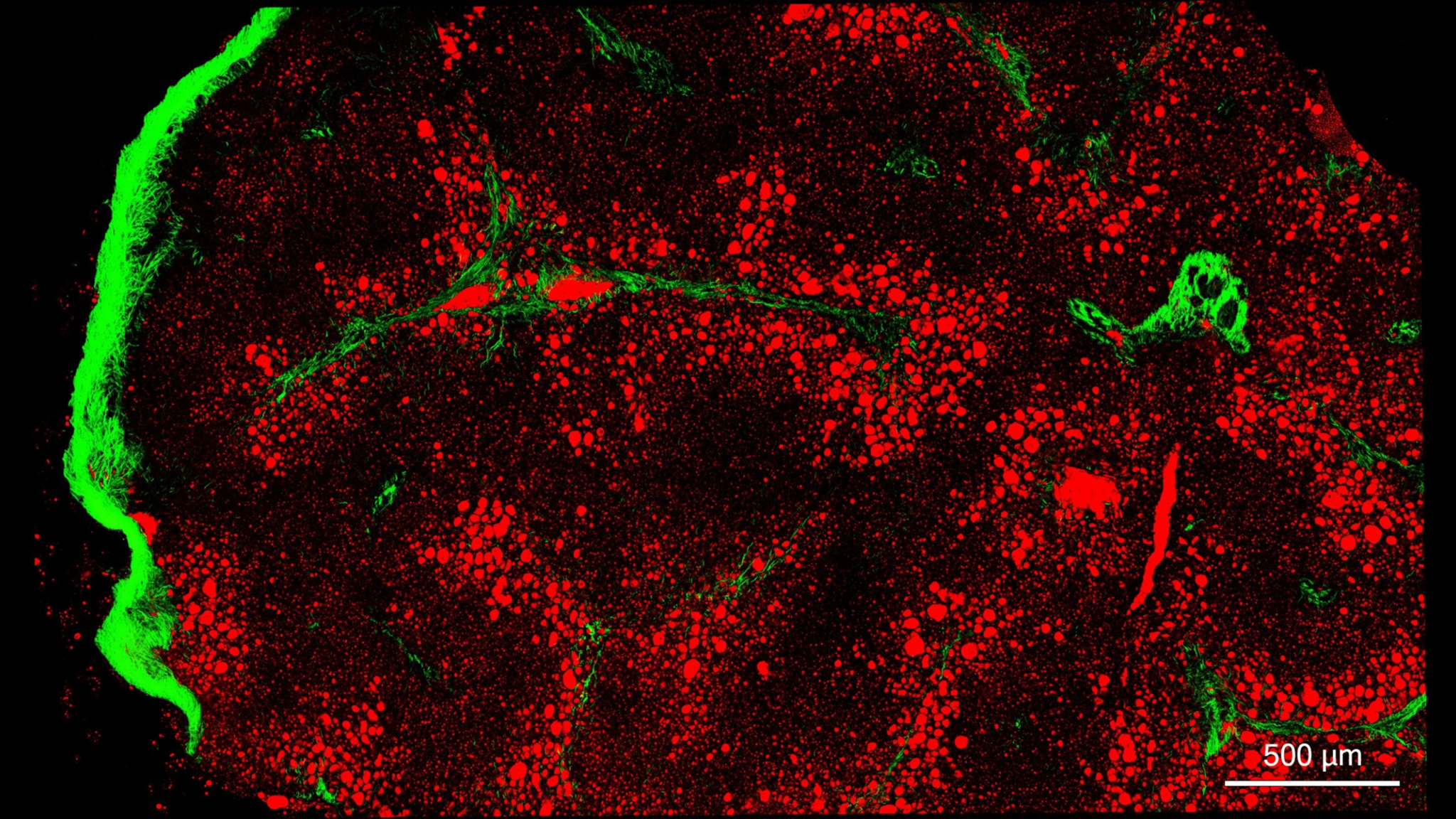
Coherent Anti-Stokes Raman Scattering

CARS microscopy is one of the label-free multiphoton imaging techniques. It allows for chemically specific imaging of biological materials without use of fluorescent labels. CARS is a three-photon nonlinear optical process where two synchronous and spatially overlapped optical beams are used to probe molecular vibrations in the sample material.

CARS microscopy is offered in Biomedicum Imaging Unit (BIU) at the University of Helsinki.
ELECTRON MICROSCOPY​

  • Immunoelectron microscopy (IEM)
  • 3D Electron Microscopy
  • Correlative light and electron microscopy (CLEM)

Immuno Electron Microscopy (IEM)

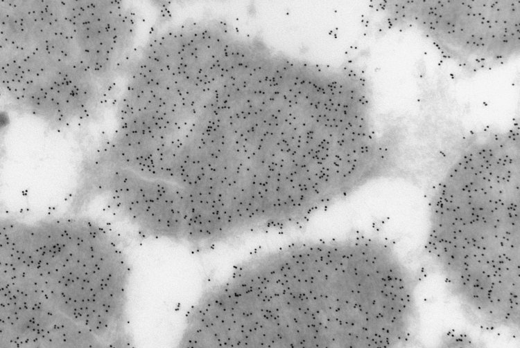
In this variation of electron microscopy (EM), specimens are chemically fixed, cryo-protected, and frozen. After this treatment, the sample is hard enough to be thin-sectioned by cryo-ultramicrotomy. The cryo-sections are then thawed and exposed to probes or antibodies. This technique is very sensitive and adapted to identifying membrane compartments in cells.

Tissue Imaging Center at Biocenter Oulu specializes in imunoelectron microscopy and provides this technology as a service.

Correlative light and electron microscopy (CLEM)

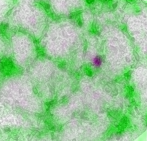
Correlative light and electron microscopy (CLEM) is the integration of two microscopy techniques, Electron Microscopy (EM) and Light Microscopy (LM), performed on the same sample and the same field of view, utilizing the strengths of each method while simultaneously reducing their individual weaknesses.

Electron Microscopy Unit at the University of Helsinki specializes in CLEM and offers it as a service to Euro-BioImaging users.

CLEM demonstration. Salo et al. 2019 Dev. Cell.

3D Electron Microscopy

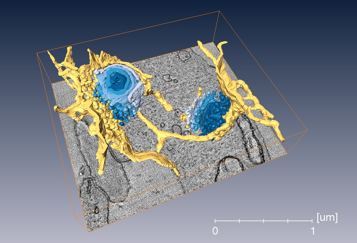
3D electron microscopy comprises a number of electron microscopy (EM) methods capable of producing 3D image data sets:

  • Electron tomography (ET): transmission electron microscopy (TEM)-based method for high-resolution imaging of relatively small areas.
  • Serial block-face scanning electron microscopy (SB-EM): for lower-resolution imaging of significantly larger areas.

Electron Microscopy Unit at the University of Helsinki specializes in 3D electron microscopy and offers it as a service to Euro-BioImaging users.

Mesoscopic Imaging

  • Optical projection tomography (OPT)
  • Selective Plane Illumination Microscopy (SPIM)
  • Multiphoton Microscopy

Optical Projection Tomography (OPT)

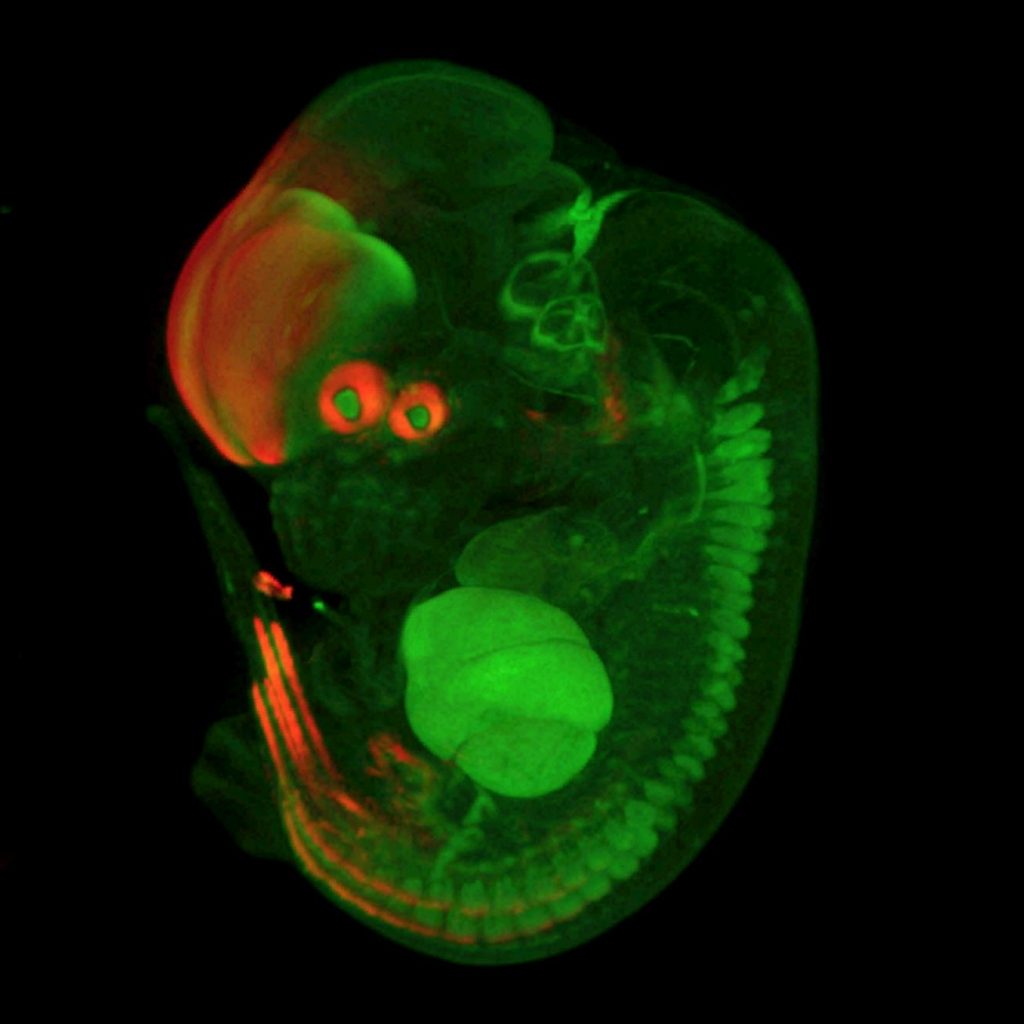
Optical Projection Tomography (OPT) is in many ways the optical equivalent of X-ray Computed Tomography (CT) or the medical CT scan. OPT differs in the way that it generally uses ultraviolet, visible, and near-infrared photons as opposed to X-ray photons. However, essential mathematics and reconstruction algorithms used for OPT and CT are similar.

Tissue Imaging Center at Biocenter Oulu specializes in imunoelectron microscopy and provides this technology as a service.

Selective Plane Illumination Microscopy (SPIM)

Single Plane Illumination Microscopy (SPIM) often referred to as light sheet fluorescence microscopy combines optical sectioning with multiple-view imaging to observe tissues and living organisms with impressive resolution. SPIM technology offers fast optical sectioning combined with minimally-invasive 3D acquisition of fluorescent specimens over time. This is achieved by focusing a thin laser light-sheet into the specimen, taking 2D images of the illuminated slice with a perpendicularly positioned detector (CCD camera). 3D stacks are obtained by moving the specimen orthogonally to the light sheet between consecutive images.

Tissue Imaging Center at Biocenter Oulu specializes in SPIM and offers this technology as a service to Euro-BioImaging users.

Super-resolution microscopy

  • Stochastic Optical Reconstruction Microscopy (STORM)
  • Stimulated Emission Depletion Microscopy (STED)

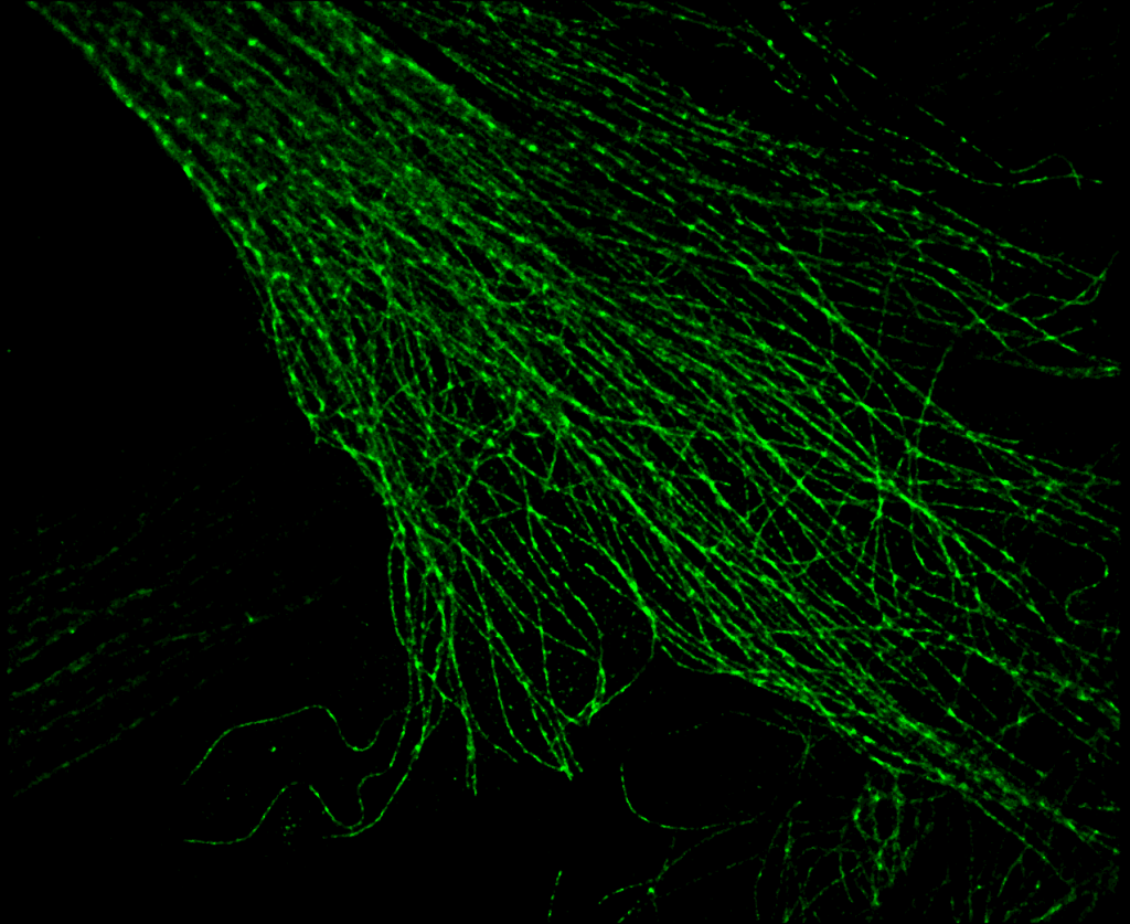
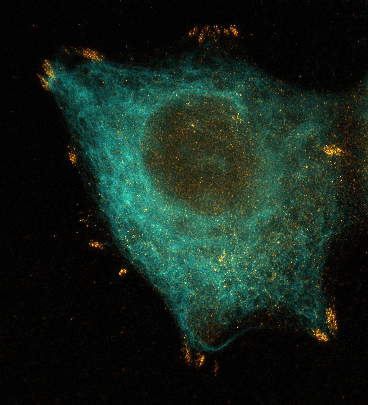
Stochastic Optical Reconstruction Microscopy (STORM)

The fundamental principle behind STochastic Optical Reconstruction Microscopy (STORM) is that the activated state of a photo-switchable molecule must lead to the consecutive emission of sufficient photons to enable precise localization before it enters a dark state or becomes deactivated by photobleaching. Additionally, the sparsely activated fluorescent molecules must be separated by a distance that exceeds the Abbe diffraction limit (in effect, greater than approximately 250 nanometers) to enable the parallel recording of many individual emitters, each having a distinct set of coordinates in the lateral image plane.

Biomedicum Imaging Unit at the University of Helsinki specializes in STORM and offers this technology as a service to Euro-BioImaging users.

Stimulated Emission Depletion Microscopy (STED)

Stimulated Emission Depletion Microscopy (STED) is a powerful super-resolution imaging modality. It creates super-resolution images by selective deactivation/depletion of fluorophores in the peripheral area, minimizing the area of illumination at the focal point and thus, enhancing the achievable resolution for a given system.

Turku BioImaging – Cell Imaging and Cytometry imaging core facility in Turku Bioscience specializes in STED microscopy and offers this technology as a service to Euro-BioImaging users.

© 2025 Finnish Advanced Microscopy Node
By Posterity Theme