NEW! Super-resolution Traction Force Microscopy for measuring mechanical forces in living cells
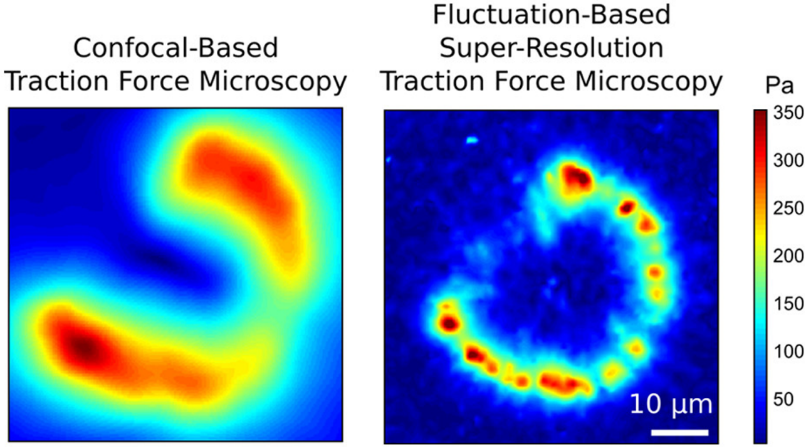
Traction Force Microscopy (TFM) has long been used to measure the forces cells exert on their environment – recent advances mean it is ready to go SUPER-RESOLUTION. Euro-BioImaging is now offering all researchers access to super-resolution Traction Force Microscopy (SR-TFM) at our Finnish Advanced Light Microscopy Node in Turku, Finland. In this interview, Dr Aki Stubb, from the University of Cambridge, explains how super-resolution Traction Force Microscopy works. Can you please briefly describe the principles behind super-resolution Traction Force Microscopy (SR-TFM)? Traction force microscopy (TFM) is a well-established technique for measuring forces exerted by the cells to surrounding extra cellular matrix. In TFM the living cells are seeded on elastic gels with known biophysical properties and that also contain embedded fluorescent beads. When the cells adhere to the gels they exert forces on it and thereby move the embedded beads. This movement can be tracked over time using confocal or…
Euro-BioImaging ERIC extends a warm welcome to its new Nodes!
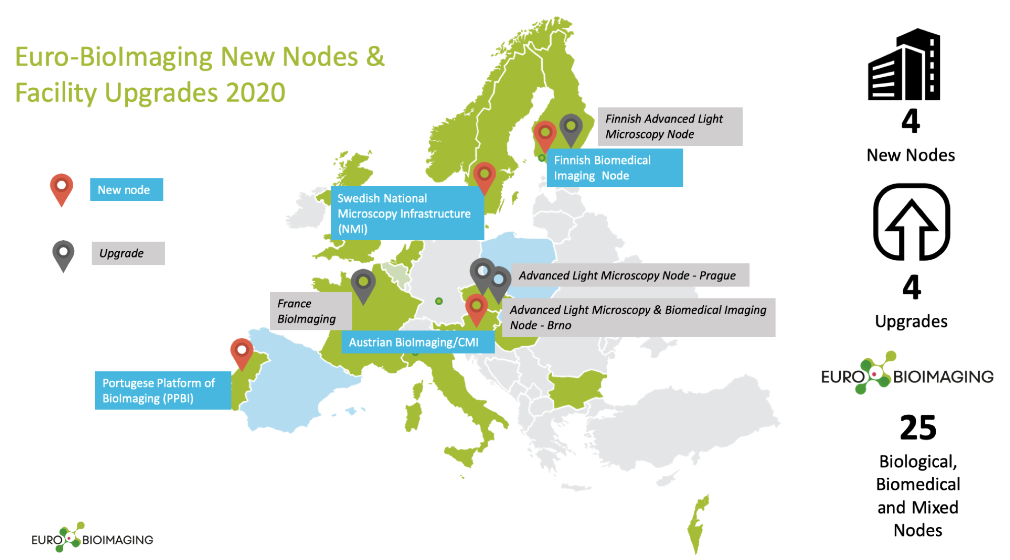
We are very pleased to announce that the Euro-BioImaging family is growing! In 2020, we welcome four new Nodes hosted by Austria, Finland, Sweden, and Portugal. These include two Biological, one Biomedical and one Mixed Node. Four existing Nodes were also granted upgrades, bringing on board new facilities and expanding their technology portfolio. A cutting-edge infrastructure These renowned imaging facilities have undergone a stringent, independent review process by the Euro-BioImaging Scientific Advisory Board (SAB) that ensures that only the most advanced imaging facilities with a cutting-edge and diverse technology offer become Euro-BioImaging Nodes. As part of the Euro-BioImaging research infrastructure, the services provided by these facilities will be open to all scientists, regardless of their discipline or affiliation. We congratulate the successful new Nodes and upgraded Nodes and look forward to sharing more news about these imaging facilities in the upcoming months. Some of the applications from this year’s Call for Nodes are still being…
Atomic Force Microscopy: Describing the mechanical properties of diverse materials
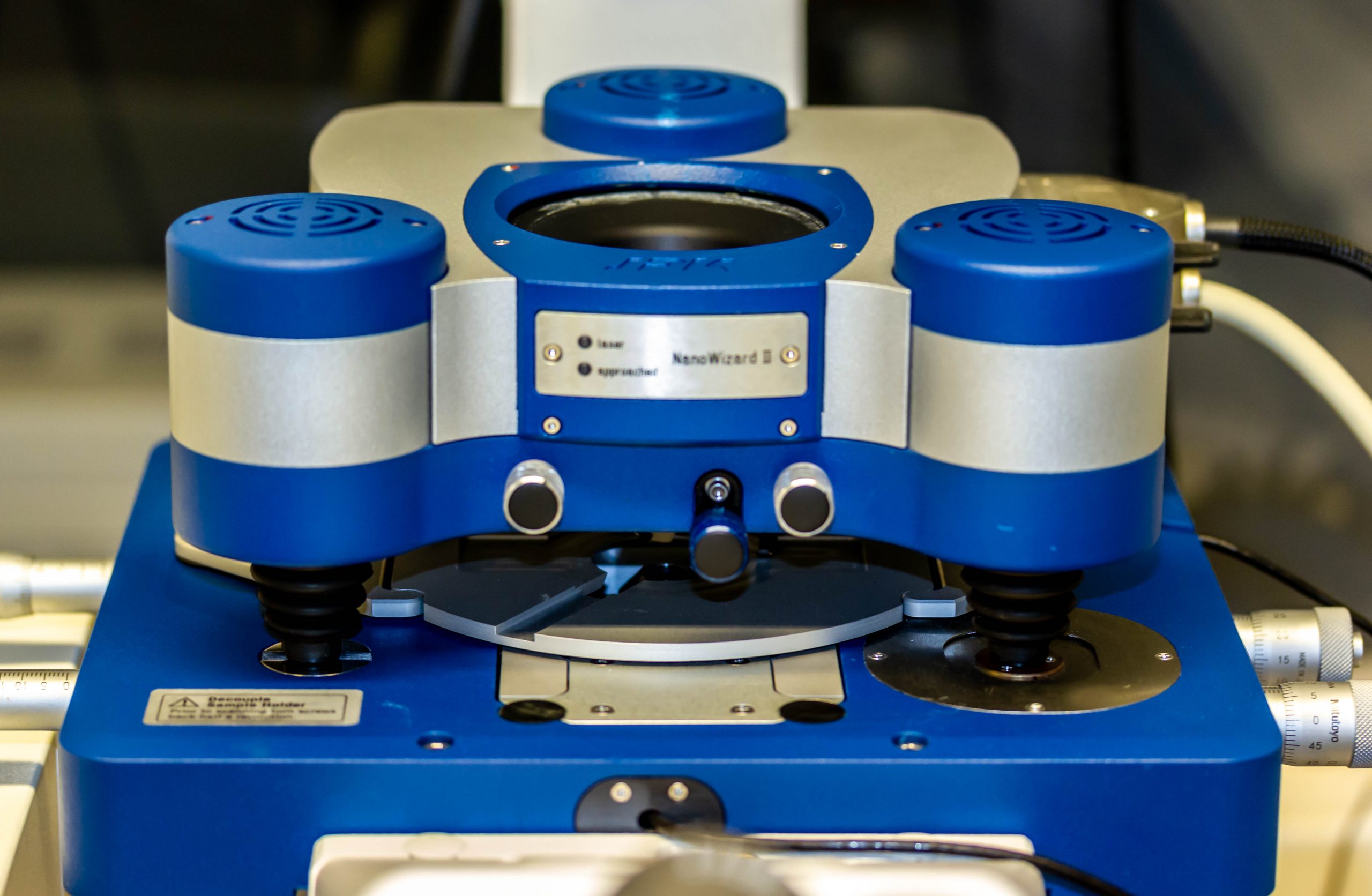
Interview with Camilo Guzmán, Manager of Euro-BioImaging’s Finnish Advanced Light Microscopy Node Please tell us who you are, where your facility is based, and what your role is. My name is Camilo Guzmán and I am the manager of the Finnish Advanced Light Microscopy Node of Euro-BioImaging. I have a background both in physics and in biology and I have been working on microscopy topics for over 17 years now. Our node has imaging facilities in Helsinki, Oulu and Turku and I am based in Turku where I take care of administrative tasks, while also providing support to different users on super-resolution microscopy, fluorescence dynamics and several biophysics methods. Read more
CARS: Label-free imaging for studying pharmaceutical materials/drug crystallization
Interview with Antti Isomäki, of the Biomedicum Imaging Unit of Helsinki BioImaging, part of Euro-BioImaging’s Finish Advanced Light Microscopy Node. Read more
New technology is offered by the Finnish Node – label-free CARS microscopy
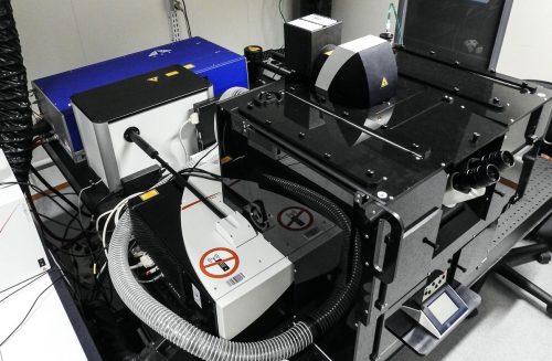
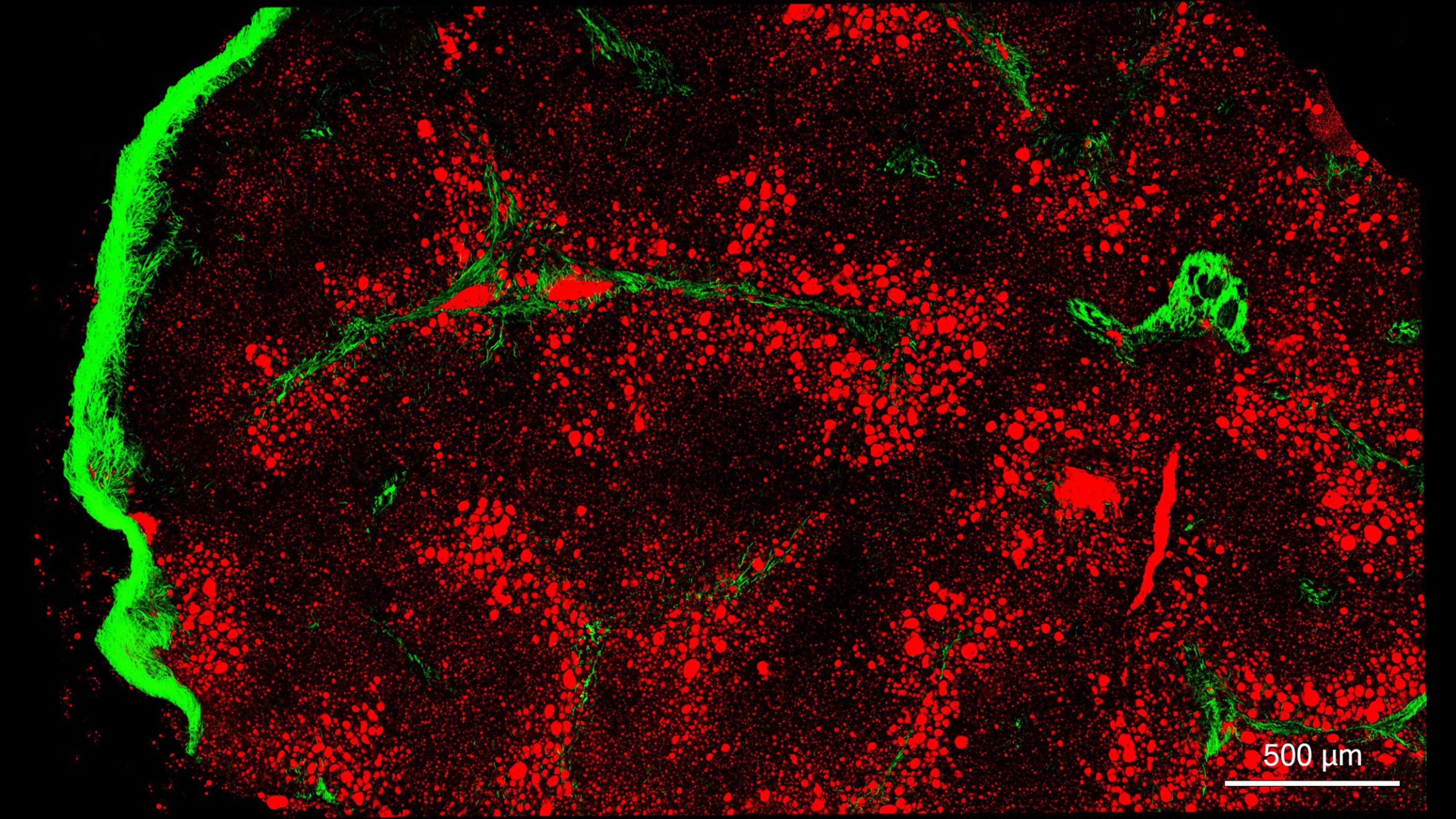
CARS microscopy is one of the label-free multiphoton imaging techniques. It allows for chemically specific imaging of biological materials without use of fluorescent labels. CARS has been proven useful in some very specific application fields. The CARS technique is particularly well suited for high-resolution label-free imaging of lipids due to the high concentration of carbon-hydrogen bonds in the lipid material. Lipid imaging has been applied to a great variety of samples including lipid droplets in fixed and live cell cultures, various tissue sections, and even small animals in vivo (e.g. zebrafish).In pharmaceutical applications, CARS microscopy is gaining more and more interest. Applications include visualization of chemical component distribution in dosage forms and drug carriers, dissolution and release, solid-state transformations during dissolution, and drug delivery into cells and tissues. You can apply to use CARS microscopy at Euro-BioImaging Web Portal.
NordForsk funding granted to Norway, Sweden, Finland, Denmark, and Iceland
NordForsk (www.nordforsk.org), an organization that facilitates research cooperation and infrastructure development in Nordic countries, has granted Bridging Nordic Microscopy Infrastructure a total of 2.5 Million NOK for 3 years. The participating countries in this consortium are Norway (coordinating Hub), Sweden, Finland, Denmark and Iceland. Bridging Nordic Microscopy Infrastructure (BNMI) is a network of national imaging infrastructures offering open access to biological imaging technologies in participating Nordic countries. BNMI partners include Norwegian Molecular Imaging Consortium (NorMIC) and Norwegian Advanced Light Microscopy Imaging Network (NALMIN), National Microscopy Infrastructure (NMI) in Sweden, Finnish Advanced Light Microscopy Node (FiALM), Danish BioImaging (DBI) Network and BioMedical Center of University of Iceland (BMC-UI). The funding will be used to strengthen international competitiveness and facilitate the development of world-leading Nordic advanced microscopy environments by supporting several levels of training, from electron and light microscopy to image analysis. Training will be delivered via scientific and technical symposia, workshops,…
Remote 3D Electron Microscopy and CLEM projects at the Finnish Node
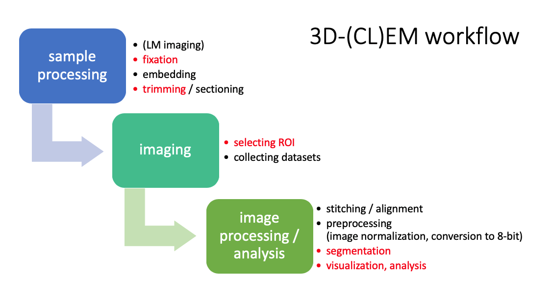
At the Finnish Node, remote access services include remote access to microscopes and image analysis workstations, virtual microscopy where a remote user joins an operator via video call, and a possibility to send samples to one of the imaging core facilities within the Finnish Node where staff perform the imaging. All of these services are possible due to ongoing developments at the Finnish Node. Eija Jokitalo, the head of Electron Microscopy Unit, at the University of Helsinki presented how imaging projects that require either 3D Electron Microscopy or CLEM (Correlative Light and Electron Microscopy) are conducted at her facility with remote users sending samples at Euro-BioImaging weekly meeting. For more information about the 3D Electron Microscopy and CLEM projects, you are welcome to read this article posted on Euro-BioImaging.
Finnish Node presents virtual microscopy at Euro-BioImaging weekly meeting
Remote access allows researchers to access instruments and analysis workstations remotely, for example, from the confines of their homes. The effects of COVID-19 pandemic, namely the temporary closure of core facilities in most of the countries or significant reduction in their activities, have accelerated the development of remote access services also at the Finnish Node. An example of remote access service being developed by the Finnish Node is virtual microscopy. It consists of an operator, generally a staff member of a core facility or an experienced researcher, acquiring image data using a microscope. A remote user joins via video call to guide an operator to image particular regions of interests. The Finnish Node presented its developments in the field of virtual microscopy at Euro-BioImaging weekly meeting on 8 May 2020. You can check a demonstration of a virtual microscopy in a short video below. https://www.youtube.com/watch?time_continue=17&v=xnPuoNTC9bI&feature=emb_titleA short video segment taken from…
Finnish Node chaired a remote access session in 2nd Euro-BioImaging Nodes meeting
2nd Euro-BioImaging Nodes meeting took place on 8 June 2020 with participants from Euro-BioImaging Nodes attending presentations and participating in discussions. This meeting took place virtually due to ongoing COVID-19 pandemic. The Finnish Node was chairing virtual access session where members of the Finnish Node presented their experiences with virtual microscopy and remote access. This session was the most popular of the three sessions during this 2nd Euro-BioImaging Nodes meeting.

