BNMI Symposium in Gothenburg, Sweden
The forth meeting of the Bridging Nordic Microscopy Infrastructures (BNMI) network will be hosted by the Centre for Cellular Imaging at the University of Gothenburg, Sweden, August 19-22, 2025. The event will bring together leading scientists from imaging research, along with key industry representatives. The primary objective of the BNMI symposium is to create an engaging and dynamic symposium that appeals to imaging researchers across disciplines. By bringing together experts from diverse imaging modalities, we aim to foster meaningful collaborations that advance the field. Whether early in your career or an experienced researcher, BNMI 2025 offers a unique opportunity to stay updated on cutting-edge imaging technologies, learn new techniques, and build valuable connections within the imaging community. The symposium will also showcase imaging research conducted by Gothenburg’s scientific community. The program will feature a compelling mix of lectures, poster sessions, and selected short talks from poster presenters, covering topics from…
Turku BioImaging is hiring image data analyst!
Turku Bioimaging (TBI) is looking for an Image Data Analyst to support cross-disciplinary projects in TBI image data team. You will– work with Finnish and international researchers and industry partners on diverse image analysis tasks– design and implement automatic image processing pipelines, test experimental image processing code, and maintain source code repositories– participate in image data management and sharing activities The position is fixed-term for the period 1.1.2025-31.12.2025 The salary for the position is set on levels 8-9 of the job demand level for other expert and support staff in the salary system of Finnish universities. In addition, a salary component based on personal work performance will be paid. Please see salary chart: https://www.sivista.fi/wp-content/uploads/2023/03/Salary-Scales-010923-and-010324.pdf If you enjoy writing code and are interested in working at the crossroads of computer science and biology, submit your application before November 4, 2024,15:00 UTC+2:00: https://abo.rekrytointi.com/paikat/index.php?jid=840&o=A_RJ&lang=en Image generated by Canva, Magic Studio
Special workshop: fast FLIM and its revolutionary applications
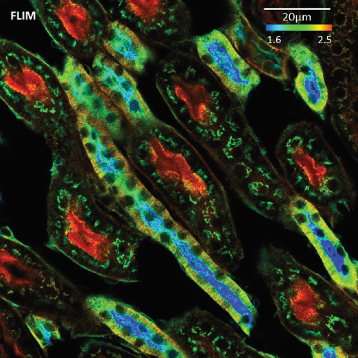
Cell Imaging and Cytometry (CIC) Core together with Immuno Diagnostic Oy is organizing a Special workshop: Fast FLIM and its revolutionary applications, which take place in BioCity on November 5th-6th, Turku, Finland. This workshop is aimed at broadly covering various new advanced applications of Fluorescence Lifetime Imaging Microscopy (FLIM) that are possible to perform on our new Leica Stellaris 8 confocal microscope at the CIC. This workshop will consist of a series of lectures and a few microscope demos on the Leica Stellaris 8 in the afternoon of Tuesday November 5th. Speakers: Register to attend the workshop by email to microscopy@bioscience.fi The lectures (as per flyer) will also be available via Zoom with link to be made available after registration.
CanSERV 3rd open call to support cancer research – apply for FREE imaging and analysis services!
We are happy to announce that the third EU-funded canSERV call is open and is now accepting applications to support cancer research projects. The researchers can apply for FREE SERVICES at several European Research Infrastructures, including Euro-BioImaging ERIC. Deadline for proposal submission is November 28, 2024, 14:00 CEST. Within this canSERV call, our Finnish Advanced Microscopy Node (FiAM) is providing open-access imaging technologies and expert services to both academic and industrial users: Some important highlights: Submission workflow: 1. Start your application by contacting the staff of the FiAM Node via contact-FiALM@eurobioimaging.fi to discuss a potential project, and inquire about imaging technologies, services, and the practicalities of the visit. In addition, visit the webpages of FiAM imaging core facilities. 2. Selection: Enter canSERV Common Access Management System, and select the services you are interested in: Service Field 2 “Advanced Technologies for Personalised Oncology” -> Service category “Imaging” -> country “Finland” -> Choose the specific service from the list (e.g.…
Practical Course on Super-Resolution Microscopy
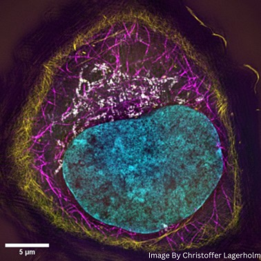
Turku Bioscience Cell Imaging and Cytometry (CIC) Core and Turku BioImaging organize the Practical Super-Resolution Microscopy course. This course will take place from Monday, September 23th to Friday, September 27th, 2024, in Turku, Finland. Every day from 9-17. The course will consist of lectures, extensive hands-on practical imaging sessions with provided samples, and group exercises. Course Description The emergence of a range of super-resolution microscopy techniques, that are capable of surpassing the classical diffraction resolution limit of about 200–250 nm in the lateral direction and 500–700 nm in the axial direction, have revolutionized light microscopy. The most commonly used super-resolution techniques are stimulated emission depletion (STED) microscopy, structural illumination microscopy (SIM), and single molecule localization microscopy (SMLM) all of which these days are widely available in commercial implementations including locally in Turku at the Turku Bioscience Cell Imaging and Cytometry (CIC) Core. In addition, recent improvements with point detector design and sensitivity has led to the…
Innovative SARS-CoV-2 Antibody Screening Technique and FAIR Data Sharing
FIMM High Content Imaging and Analysis (FIMM-HCA) team has been recently published the work on mini-immunofluorescence assay to test for SARS-CoV-2 antibodies in patient blood samples in Cell Report Methods journal: https://doi.org/10.1016/j.crmeth.2023.100565 Researchers from the FIMM High Content Imaging and Analysis (FIMM-HCA) Unit (partner of the Finnish Advanced Microscopy Node) have unveiled a cutting-edge method combining image-based serology and machine learning to detect SARS-CoV-2 antibodies. Led by Lassi Paavolainen, Academy Research Fellow at the Institute for Molecular Medicine Finland, their mini-immunofluorescence assay leverages custom neural network analysis for high-throughput testing in low-biosafety environments. This breakthrough not only enhances disease detection but also sets a high standard in data accessibility through the BY-COVID project. The entire dataset, including raw images and segmentation masks, is now publicly available on the BioImage Archive, marking a pivotal move towards FAIR (Findable, Accessible, Interoperable, Reusable) data sharing. The metadata for the images is described according…
Visiting Professor Teng-Leong Chew in Turku – InFLAMES minisymposium and one-day microscopy workshop
Great news! Professor Teng-Leon Chew from Advanced Imaging Center, HHMI´s Janelia Research Campus will visit Turku next week! Professor Chew will give a talk at InFLAMES minisymposium and lead a one-day microscopy 🔬 workshop! ▶ InFLAMES Visiting Professor minisymposium💡 Topic: “Microscopy technologies: today and tomorrow”, Professor Teng-Leong Chew, Director of Advanced Imaging Center, Janelia Research Campus, USA📅 Monday, June 17, 2024, 12-14📍 Lauren 1, Medisiina D, Turku, Finland🔗 More info: https://inflames.utu.fi/events/inflames-minisymposium-teng-leong-chew-and-pekka-lappalainen/ ▶ InFLAMES mini microscopy workshop💡 “Experimental design for hypothesis-driven quantitative fluorescence microscopy” by Professor Teng-Leong Chew📅 Tuesday, June 18, 2024, 9-16📍 Lauren 2, Medisiina D, Turku, Finland🔗 More info: https://inflames.utu.fi/events/52015/ Topics covered: No previous knowledge of imaging techniques or microscopy is required, but even more experienced researches are sure to learn something new! Register now using the link: webropol.com/s/leong-miniworkshop
A Guide to FAIR Bioimage Data 2024
Do you perform biological imaging and want to maximise the potential of your bioimaging data? Euro-BioImaging is organizing free online workshop, “Euro-BioImaging’s Guide to FAIR Bioimage Data” on Thursday, 23 May 2024, from 14:00 to 17:00 CEST. Everyone is welcome! In this interactive online workshop you will learn about the FAIR principles in the context of bioimaging data. Designed for researchers across all scales of bioimaging, from molecules to humans, this workshop will provide simple yet effective steps for a smooth start to your FAIR journey. You will get to now about the benefits of FAIR data for molecular, cellular as well as pre-clinical imaging and best practices for data management. Full programme here: https://www.eurobioimaging.eu/upload/Schedule_fairguide.pdf Register here: https://us02web.zoom.us/meeting/register/tZ0ldu2grz4pGdyUhJJgOLpml1elNcd1-Iyx Time & Date: 14:00-17:00 CEST, May 23rd-24th, 2024.
CanSERV 2nd open call to support cancer research – apply for FREE imaging and analysis services!
We are happy to announce that the second EU-funded canSERV call is open and is now accepting applications to support cancer research projects. The researchers can apply for FREE SERVICES at several European Research Infrastructures, including Euro-BioImaging ERIC. Deadline for proposal submission is May 21st, 2024, 14:00 CEST. Within this canSERV call, our Finnish Advanced Microscopy Node (FiAM) is providing open-access imaging technologies and expert services to both academic and industrial users: Some important highlights: Submission workflow: 1. Start your application by contacting the staff of the FiAM Node via contact-FiALM@eurobioimaging.fi to discuss a potential project, and inquire about imaging technologies, services, and the practicalities of the visit. In addition, visit the webpages of FiAM imaging core facilities. 2. Selection: Enter canSERV Common Access Management System, and select the services you are interested in: Service Field 2 “Advanced Technologies for Personalised Oncology” -> Service category “Imaging” -> country “Finland” -> Choose the specific service from the list (e.g.…
BNMI symposium 2024 in Norway
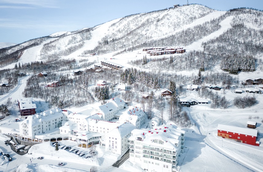
The third meeting of the Bridging Nordic Microscopy Infrastructures (BNMI) network will take place at the Dr. Holms Hotel, Geilo, Norway at April 9-12, 2024. The goal of the BNMI network symposium is to bring the Nordic imaging infrastructures closer together, to strengthen our collaboration and to encourage networking between application scientists, developers and industry in order to improve the quality and impact of microscopy in the Nordic countries . With this meeting the BNMI aim to encourage interactions among the different Nordic imaging facilities and create interactive networks in life science imaging. BNMI offers an exciting scientific program as well as social program. The social program will include cross-country skiing, alpine skiing, bowling, shuffleboard, spa and if the weather allows it, outdoor Après–ski. List of exciting topics: The invited distinguished speakers are: Venue: Dr. Holms Hotel, Geilo, Norway Dates: April 9/10-12, 2024 Registration deadline: February 8, 2024 Abstract submission…


