35th BioCity Symposium: VISION, EXPANDED
Turku is the beating heart of European imaging! The 35th BioCity Symposium “Vision, Expanded” brings together leading minds from academia and industry to explore imaging’s central role in life science discovery. The Symposium will spotlight cutting-edge advances in light microscopy, PET imaging, and AI-powered image analysis, while fostering dialogue on data integration, emerging scientific applications, and translational potential. Expect high-level talks and vibrant discussion that push the boundaries of imaging science. Original artwork by Joanna Pylvänäinen, 2025 Registration and abstract submission is now open, click! Submit your abstract at latest Friday 5 June 2026, there is quota of 125. First come first serve! Register to the event at latest Friday 14 August 2026 Invited Speakers Sally Eldeghaidy, University of Nottingham, UKRicardo Henriques, ITQB NOVA, PortugalHarry Hu, Mayo Clinic Jacksonville, Florida, USATeemu Ihalainen, Tampere University, FinlandSinem Karaman, University of Helsinki, FinlandKarolina Punovuori, MultivisionDx Oy; University of Helsinki, FinlandJörg Renkawitz, Ludwig-Maximilians-Universität München,…
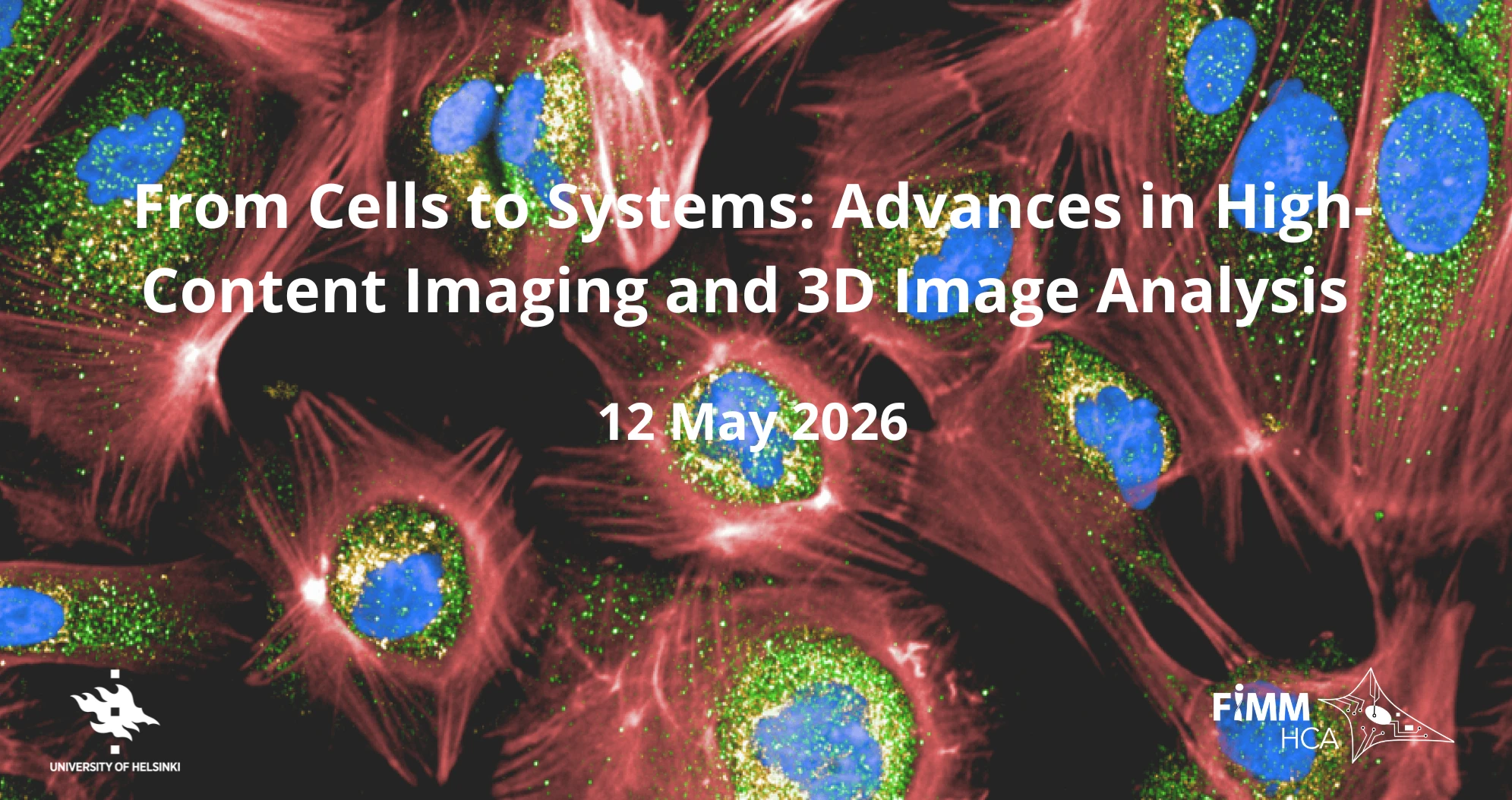
From Cells to Systems: Advances in High-Content Imaging and 3D Image Analysis symposium in Helsinki
FIMM High Content Imaging and Analysis unit is organizing symposium “From Cells to Systems: Advances in High-Content Imaging and 3D Image Analysis” May 12, 2026 in Helsinki, Finland.🗓️ May 12, 2026🕰️ 09-17📍 Biomedicum 1 | Helsinki | Finland Program Talks in three sessions by international and local speakers showcasing recent advances in: Session I: 3D Imaging and analysis in complex systems:· Peter Horvath, Helmholtz Munich, AI4Health Institute, Germany; Director; Institute of Biochemistry, Biological Research Centre (BRC), Hungary· Heidi Haikala, PI/Ass. Prof., Faculty of Medicine and FIMM-HiLIFE, University of Helsinki & co-founder of SOLID IO, Finland· Lassi Paavolainen, PI, Institute for Molecular Medicine Finland-FIMM, HiLIFE, University of Helsinki, Finland Session II: High-content drug screening & phenotypic discoveries in disease:· Jordi Carreras-Puigvert, Ass. Prof., Department of Pharmaceutical Biosciences, Uppsala University & CSO & Co-Founder at Pixl Bio, Sweden· Simon Pfisterer, PI, Faculty of Medicine, University of Helsinki, & CSO, Founder, MONCYTE health Ltd., Finland Session III: Cell Painting Assay Technology and Integrative Omics:· Helena Kilpinen, PI, Ass. Prof., Institute for Molecular Medicine Finland-FIMM, HiLIFE;…
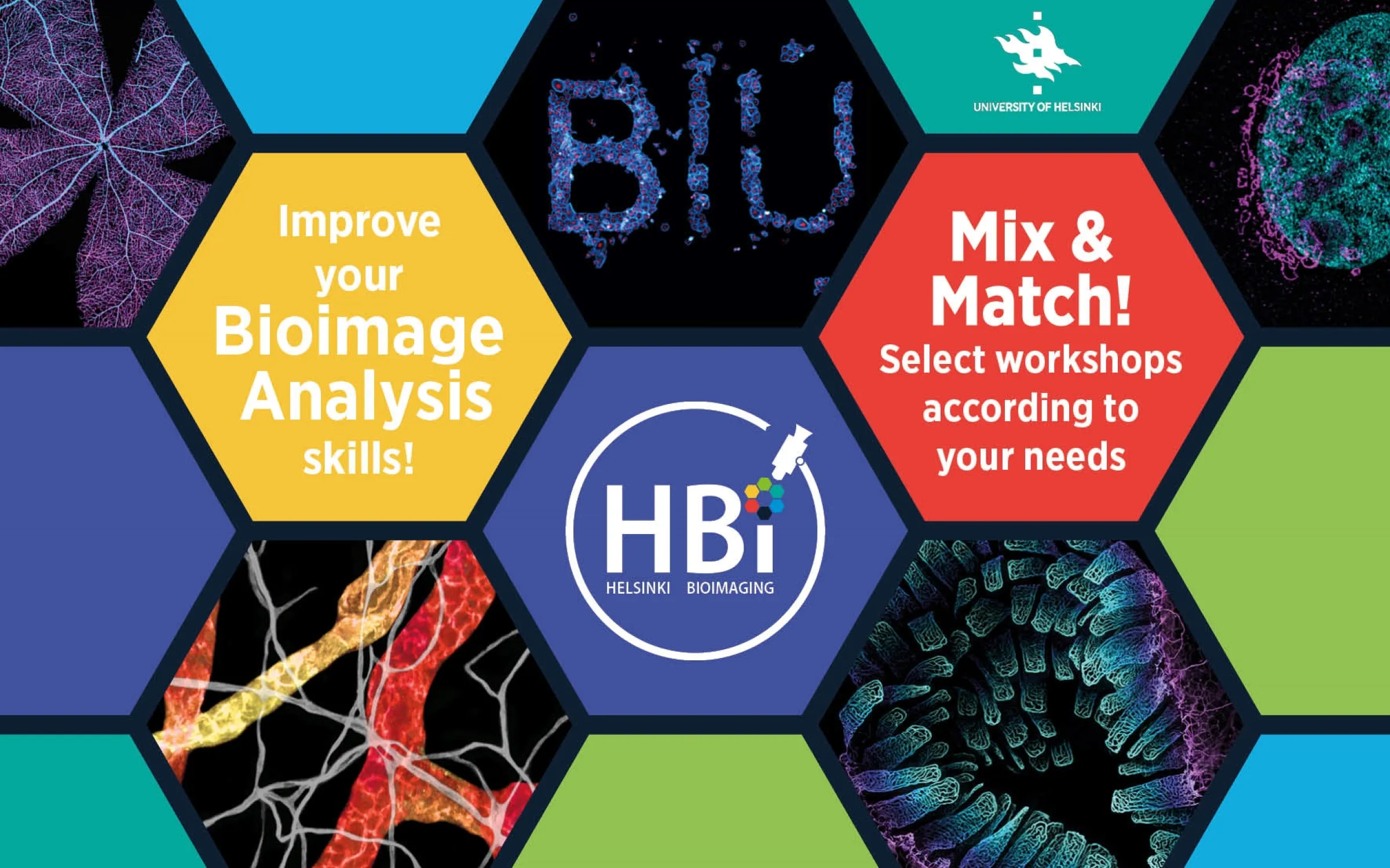
Bioimage Analysis Course 2026 organized by Helsinki BioImaging
Biomedicum Imaging Unit, Helsinki BioImaging, organizes “Bioimage Analysis Course” in May 2026. When: On-site lectures on 04.05., 06.05.2026, and hands-on workshops on 5-13.05.2026 Where: Biomedicum, Haartmaninkatu 8 & Haartman Institute, Haartmaninkatu 3, Helsinki, Finland (Image: Images: Satu Paavonsalo, Sinem Karaman and Kirstin Vonderstein – Graphics: Kirstin Vonderstein) Lectures Monday 4th May 2026 Wednesday 6th May 2026 Confirmed Speakers Hands-on Workshops (Mix and Match!) Apply for the workshops you like and we will send you your individual schedule.The workshops will give you the opportunity to get your hands on a wide range of image‑analysis tools. All the software will be introduced is either free & open‑source or available for you to use on the BIU image‑analysis workstations: Registartion Register by 29.03.2026: For PhD students that want to earn 1ECTS: Registration for the whole course (all lectures + 3 hands-on workshops) For every one else: Mix and match as you like! Registration…
SCANDEM2026 in Oulu
SCANDEM2026, the Annual Meeting of Nordic Microscopy Society, will take place in Oulu, Finland, June 9-12, 2026. The researchers, professionals, and students from across the Nordic countries will come to explore the latest developments in microscopy and imaging in the fields of life and materials sciences. This meeting offers an excellent opportunity for researchers and students to: Venue: Biocenter Oulu, Kieppi building, Aapistie 5A, Oulu, Finland Registration deadline: April 15, 2026 Abstract submission by March 31, 2026 (for poster and selected talks) The meeting is organized in collaboration with the University of Oulu and the Nordic Microscopy Society. More information via this link
BNMI Symposium in Levi, Finland
The 5th meeting of the Bridging Nordic Microscopy Infrastructures (BNMI) network will be hosted by the Finnish Advanced Microscopy Node at the Levi Summit Event Centre & Hotel Levi Panorama, Lapland, Finland, November 23/24-27, 2026. The goal of the BNMI symposium is to bring the Nordic imaging infrastructures, leading imaging scientists, and industry representatives together for insightful discussions and collaboration to improve the quality and impact of microscopy in the Nordic countries. By bringing together experts from different imaging modalities and career stages, we aim to foster meaningful collaborations and offer a unique opportunity to stay updated on cutting-edge imaging technologies, learn new techniques, and build valuable connections within the imaging community. BNMI2026 webpage: https://eurobioimaging.fi/FiAM/bnmi2026/ Program highlights We will offer an exciting scientific program, which will consist of industry exhibitions, workshops, scientific talks, poster sessions, and panel discussion. Topics that will be covered include: The social program will include cross-country…
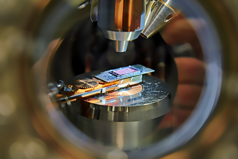
Turku BioImaging is hiring a Senior Laboratory Engineer in Advanced Imaging
The Advanced Imaging core facility in Turku (AIC-Turku, previously CIC) is seeking a highly experienced and motivated Laboratory Engineer (Senior) in Advanced Imaging. The employment is fulltime and for the period 1.5.2026-31.12.2028. Image Source: Åbo Akademi University image bank More information about the imaging facility is here. Work tasks As a Laboratory Engineer (Senior) in Advanced Imaging, you will support cell and tissue imaging needs across research areas including cancer, neurobiology, and immunology. You will work with advanced light microscopy, biological image processing methods, and imaging data analysis. You will also play a key role in the continuous development of AIC-Turku services and operations. Your responsibilities will include: Qualifications We are primarily looking for a Laboratory Engineer (Senior) with more than five year’s experience of working in a microscopy facility or in a company as a microscopy application specialist, having also gained some experience in leading processes (leadership). An applicant…
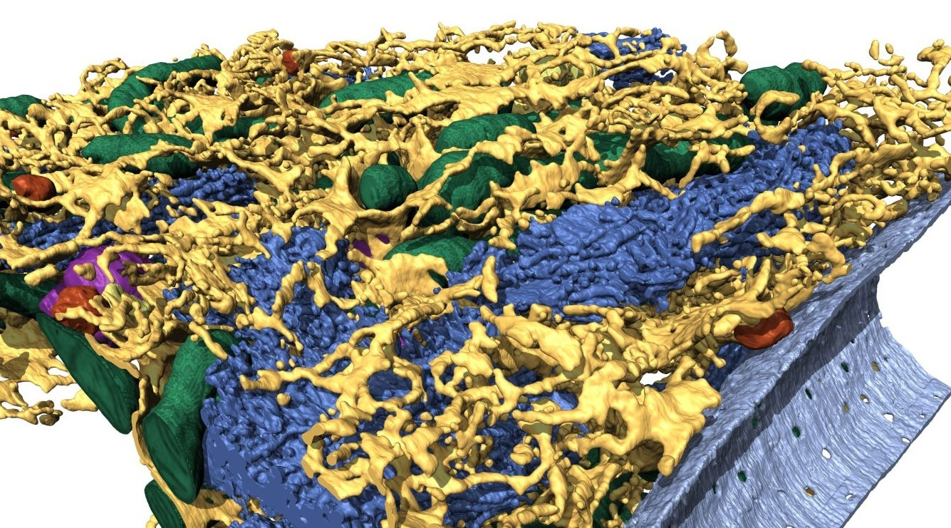
Frontiers in Imaging: celebrating the EMBI-ZEISS partnership on volumeEM
The Electron Microscopy Unit (EMBI), Helsinki BioImaging and ZEISS Microscopy are organizing a symposium “Frontiers in Imaging: celebrating the EMBI-ZEISS partnership on volumeEM” to celebrate the inauguration of EMBI new microscopes and the Labs@location partnership with Zeiss. VolumeEM technology unveils the ultrastructure of biological samples in 3D, capturing details from micrometer to millimeter volumes at nanometer resolution. Discover the diverse methods that enable successful volumetric imaging of cells and tissues, and learn how to analyze these results effectively. Dates: December 2-3, 2025 Location: EMBI, Institute of Biotechnology, Viikinkaari 9, University of Helsinki, 00790, Helsinki The program consists of lectures, a visit to EMBI and workshops (SBF-SEM, FIB-SEM, Micro-CT, Image analysis). Program an registration via this link.
Winners of the “Summer” Euro-BioImaging imaging contest are announced
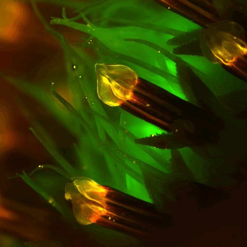
Euro-BioImaging announced the “Summer” round of the Four Seasons of the Invisible Imaging Contest in early 2025. The competition highlights seasonally inspired imaging work that reveals the unseen beauty of the microscopic world. FiAM Node is delighted to share that Kirstin Vonderstein, PhD from the Biomedicum Imaging Unit, the University of Helsinki, received second place in this competition for the image Summer’s Hidden Glow: “Autofluorescence of Cosmos Bipinnatus“. This image captures the stamens of a Cosmos bipinnatus flower that was found blooming along the wayside during the height of summer. Stereomicroscope image (1.63× magnification) of an unstained Cosmos bipinnatus flower, showing natural autofluorescence in yellow, red, and green. Imaging was performed at the Biomedicum Imaging Unit, the University of Helsinki. Kirstin Vonderstein will receive reimbursement of up to €500 to attend a scientific conference. The “Autumn” round of the Four Seasons of the Invisible Imaging Contest is open, and the submission deadline is December…
Helsinki BioImaging is hiring image analysis specialist!
Helsinki Bioimaging (HBI) is seeking a highly motivated and goal-oriented Image Analysis Specialist/Engineer/Coordinator to develop and apply cutting-edge light microscopy image analysis workflows. Image generated by Canva AI, Magic Studio The position This role offers a unique opportunity to apply and integrate advanced image processing techniques to progress the fields of biomedical and life sciences. Working within a team of biologists, physicists and engineers, the specialist will help build robust and reproducible protocols to extract meaningful quantitative information from both simple and complex microscopy datasets and present it effectively. This is a joint position between Biomedicum Imaging Unit (BIU) at the Meilahti campus and Light Microscopy Unit (LMU) at the Viikki campus, which are open access core facilities offering a wide variety of basic and advanced light microscopy techniques to several hundred local, national and international users annually. Specific tasks Qualifications In addition, one or more of the following would…
Åbo Akademi is hiring a university researcher in bioimaging
Åbo Akademi University is looking for a University Researcher in Bioimaging for the period 1 November 2025 until 31 December 2030. The appointment is full-time and located at the Biochemistry and Cell Biology unit at the Faculty of Science and Engineering in Turku, Finland. The successful candidate will be an expert in advanced light microscopy and/or image analysis, with research interests closely aligned with those of existing research groups within Cell Biology. Image source: Åbo Akademi University image bank The tasks are specified below: Research (70%) Teaching (30%) Coordinate advanced-level courses (Master’s and PhD level) in: The position is fixed-term for the period 1.11.2025-31.12.2030 The university researcher’s salary is based on levels 6-7 of the job requirement levels for teaching and research staff. In addition, an individual salary component based on personal work performance is added to the salary. Please see the salary chart for teaching and research personnel. More information…