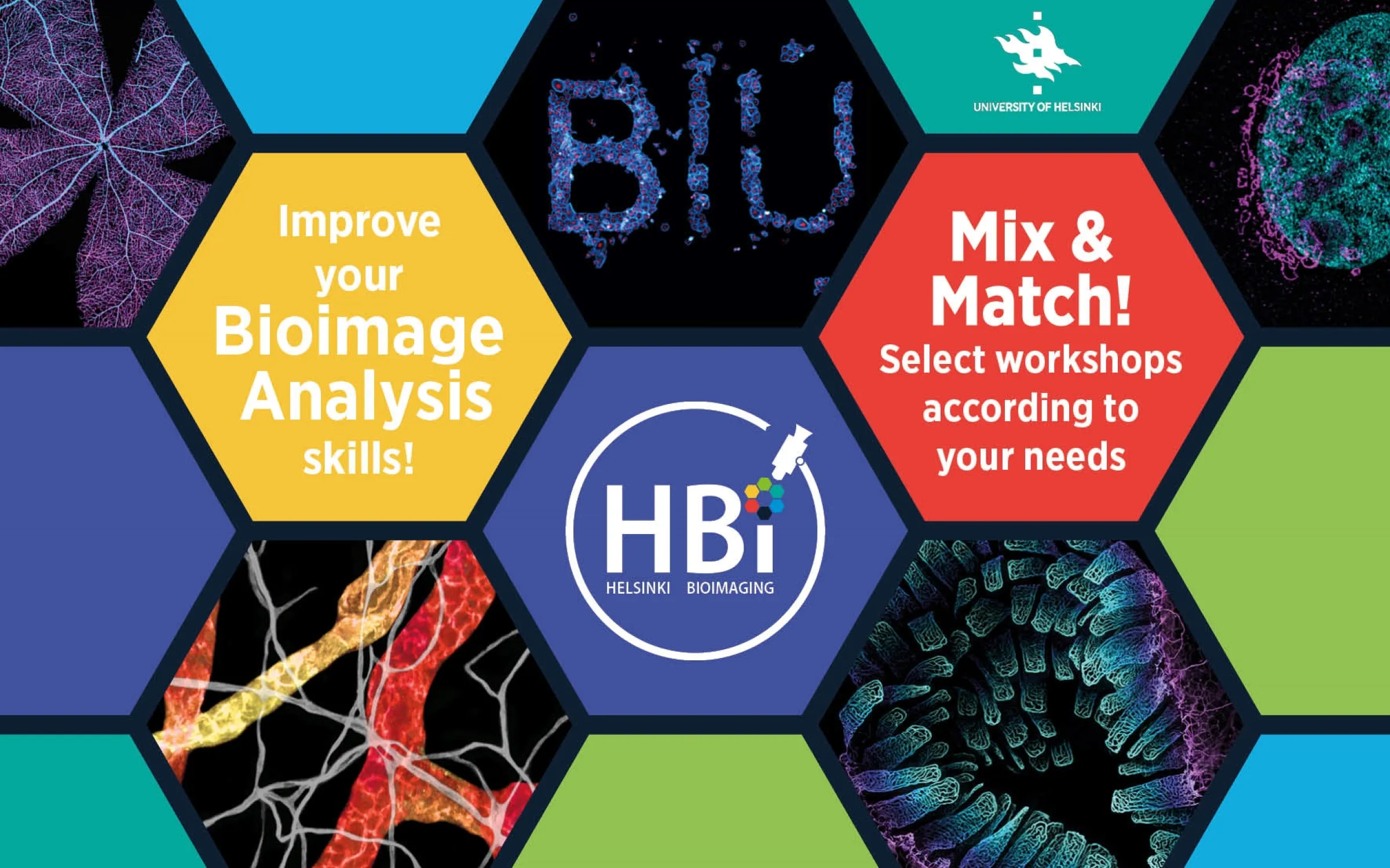
Bioimage Analysis Course 2026 organized by Helsinki BioImaging
Biomedicum Imaging Unit, Helsinki BioImaging, organizes “Bioimage Analysis Course” in May 2026. When: On-site lectures on 04.05., 06.05.2026, and hands-on workshops on 5-13.05.2026 Where: Biomedicum, Haartmaninkatu 8 & Haartman Institute, Haartmaninkatu 3, Helsinki, Finland (Image: Images: Satu Paavonsalo, Sinem Karaman and Kirstin Vonderstein – Graphics: Kirstin Vonderstein) Lectures Monday 4th May 2026 Wednesday 6th May 2026 Confirmed Speakers Hands-on Workshops (Mix and Match!) Apply for the workshops you like and we will send you your individual schedule.The workshops will give you the opportunity to get your hands on a wide range of image‑analysis tools. All the software will be introduced is either free & open‑source or available for you to use on the BIU image‑analysis workstations: Registartion Register by 29.03.2026: For PhD students that want to earn 1ECTS: Registration for the whole course (all lectures + 3 hands-on workshops) For every one else: Mix and match as you like! Registration…
Practical Course on Super-Resolution Microscopy
Turku Bioscience Cell Imaging and Cytometry (CIC) Core and Turku BioImaging organize the Practical Super-Resolution Microscopy course. This course will take place from Monday, September 23th to Friday, September 27th, 2024, in Turku, Finland. Every day from 9-17. The course will consist of lectures, extensive hands-on practical imaging sessions with provided samples, and group exercises. Course Description The emergence of a range of super-resolution microscopy techniques, that are capable of surpassing the classical diffraction resolution limit of about 200–250 nm in the lateral direction and 500–700 nm in the axial direction, have revolutionized light microscopy. The most commonly used super-resolution techniques are stimulated emission depletion (STED) microscopy, structural illumination microscopy (SIM), and single molecule localization microscopy (SMLM) all of which these days are widely available in commercial implementations including locally in Turku at the Turku Bioscience Cell Imaging and Cytometry (CIC) Core. In addition, recent improvements with point detector design and sensitivity has led to the…
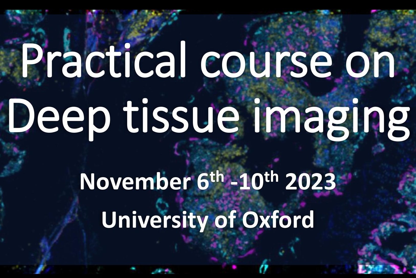
Practical course on Deep tissue imaging, Oxford, UK
Turku Cell Imaging and Cytometry Core Facility has teamed up with the Oxford-Zeiss Centre of Excellence at the Kennedy Institute of Rheumatology at the University of Oxford to organize a joint “Practical Course on Deep Tissue Imaging”. This course will take place in Oxford, UK on November 6-10. Course Description:Deep imaging (>50 μm) in biomedical samples such as thick tissue sections, spheroids, and organoids with fluorescence microscopy has become a much more common quest by bio-medical researchers. Yet, such imaging is very challenging as a consequence of difficulties with deep penetrative staining, and with optical limitations including light penetration, light scattering, and refractive index mismatch. The aim of this course is to collectively explore the use of a range of state-of-the-art fluorescence microscopes that are available within the Oxford – ZEISS Centre of Excellence at the University of Oxford, in order to gain a better understanding of the advantages and…