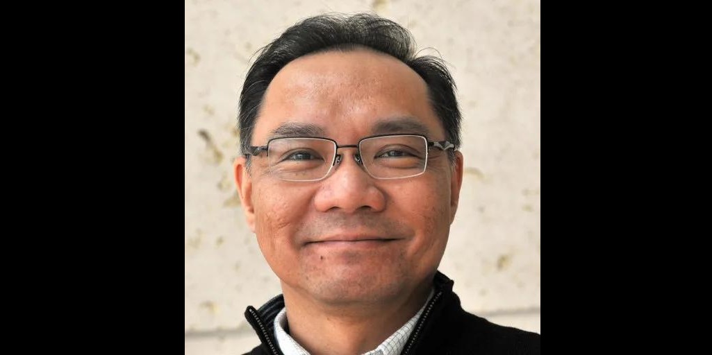
Visiting Professor Teng-Leong Chew in Turku – InFLAMES minisymposium and one-day microscopy workshop
Great news! Professor Teng-Leon Chew from Advanced Imaging Center, HHMI´s Janelia Research Campus will visit Turku next week! Professor Chew will give a talk at InFLAMES minisymposium and lead a one-day microscopy 🔬 workshop! ▶ InFLAMES Visiting Professor minisymposium💡 Topic: “Microscopy technologies: today and tomorrow”, Professor Teng-Leong Chew, Director of Advanced Imaging Center, Janelia Research Campus, USA📅 Monday, June 17, 2024, 12-14📍 Lauren 1, Medisiina D, Turku, Finland🔗 More info: https://inflames.utu.fi/events/inflames-minisymposium-teng-leong-chew-and-pekka-lappalainen/ ▶ InFLAMES mini microscopy workshop💡 “Experimental design for hypothesis-driven quantitative fluorescence microscopy” by Professor Teng-Leong Chew📅 Tuesday, June 18, 2024, 9-16📍 Lauren 2, Medisiina D, Turku, Finland🔗 More info: https://inflames.utu.fi/events/52015/ Topics covered: No previous knowledge of imaging techniques or microscopy is required, but even more experienced researches are sure to learn something new! Register now using the link: webropol.com/s/leong-miniworkshop

A Guide to FAIR Bioimage Data 2024
Do you perform biological imaging and want to maximise the potential of your bioimaging data? Euro-BioImaging is organizing free online workshop, “Euro-BioImaging’s Guide to FAIR Bioimage Data” on Thursday, 23 May 2024, from 14:00 to 17:00 CEST. Everyone is welcome! In this interactive online workshop you will learn about the FAIR principles in the context of bioimaging data. Designed for researchers across all scales of bioimaging, from molecules to humans, this workshop will provide simple yet effective steps for a smooth start to your FAIR journey. You will get to now about the benefits of FAIR data for molecular, cellular as well as pre-clinical imaging and best practices for data management. Full programme here: https://www.eurobioimaging.eu/upload/Schedule_fairguide.pdf Register here: https://us02web.zoom.us/meeting/register/tZ0ldu2grz4pGdyUhJJgOLpml1elNcd1-Iyx Time & Date: 14:00-17:00 CEST, May 23rd-24th, 2024.
CanSERV 2nd open call to support cancer research – apply for FREE imaging and analysis services!
We are happy to announce that the second EU-funded canSERV call is open and is now accepting applications to support cancer research projects. The researchers can apply for FREE SERVICES at several European Research Infrastructures, including Euro-BioImaging ERIC. Deadline for proposal submission is May 21st, 2024, 14:00 CEST. Within this canSERV call, our Finnish Advanced Microscopy Node (FiAM) is providing open-access imaging technologies and expert services to both academic and industrial users: Some important highlights: Submission workflow: 1. Start your application by contacting the staff of the FiAM Node via contact-FiALM@eurobioimaging.fi to discuss a potential project, and inquire about imaging technologies, services, and the practicalities of the visit. In addition, visit the webpages of FiAM imaging core facilities. 2. Selection: Enter canSERV Common Access Management System, and select the services you are interested in: Service Field 2 “Advanced Technologies for Personalised Oncology” -> Service category “Imaging” -> country “Finland” -> Choose the specific service from the list (e.g.…
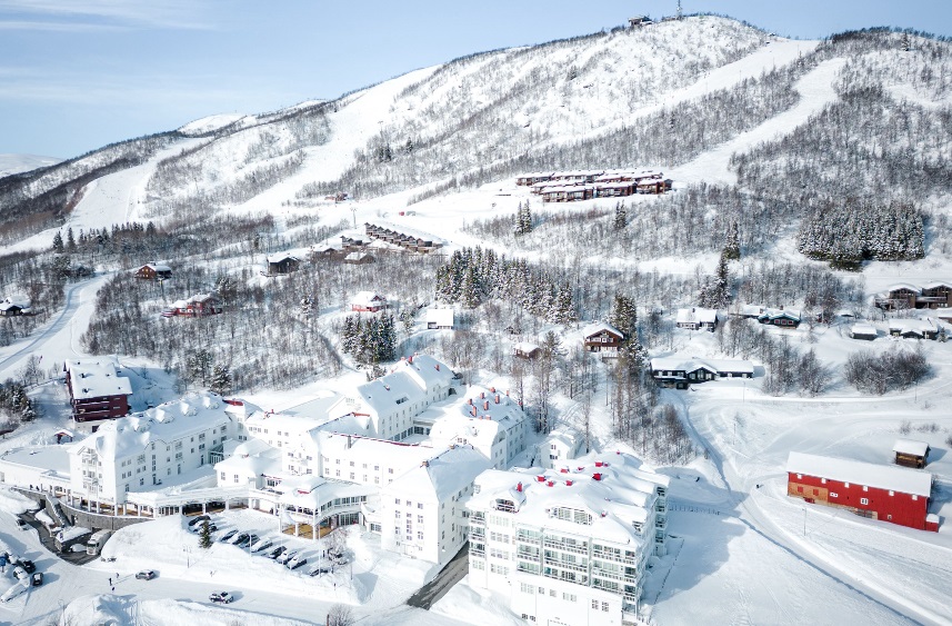
BNMI symposium 2024 in Norway
The third meeting of the Bridging Nordic Microscopy Infrastructures (BNMI) network will take place at the Dr. Holms Hotel, Geilo, Norway at April 9-12, 2024. The goal of the BNMI network symposium is to bring the Nordic imaging infrastructures closer together, to strengthen our collaboration and to encourage networking between application scientists, developers and industry in order to improve the quality and impact of microscopy in the Nordic countries . With this meeting the BNMI aim to encourage interactions among the different Nordic imaging facilities and create interactive networks in life science imaging. BNMI offers an exciting scientific program as well as social program. The social program will include cross-country skiing, alpine skiing, bowling, shuffleboard, spa and if the weather allows it, outdoor Après–ski. List of exciting topics: The invited distinguished speakers are: Venue: Dr. Holms Hotel, Geilo, Norway Dates: April 9/10-12, 2024 Registration deadline: February 8, 2024 Abstract submission…
Academy of Finland grants FIRI funding to the Finnish Advanced Microscopy Node
Finnish Advance Microscopy (FiAM) Node received the Academy of Finland “FIRI 2023: roadmap research infrastructures 2021–2024” funding. A total of 7,2 million euros will be granted to the FiAM Node over the period of 2024-2026. Decision can be found here. The FIRI funding will be used to strength and upgrade the technological capabilities and services of imaging core facilities that are part of the FiAM Node in Helsinki (University of Helsinki), Turku (University of Turku & Åbo Akademi University), and Oulu (University of Oulu). FiAM Node led by Turku BioImaging is one of the most popular service unit of the large European landmark infrastructure Euro-BioImaging. FiAM’s services are very important also for numerous Finnish health and drug development companies. With the FIRI funding FiAM Node will establish an extensive set of the newest microscopy methods, capable of high-resolution 3D imaging also from living samples. FiAM will also establish 3D image…
CanSERV call to support cancer research
We are happy to announce that the first EU-funded canSERV call is open and is now accepting applications to support cancer research projects. The researchers can apply for FREE SERVICES at several European Research Infrastructures, including Euro-BioImaging ERIC. Deadline for proposal submission is January 4th, 2024. Within this canSERV call, our Finnish Advanced Microscopy Node (FiAM) is providing open-access imaging technologies and expert services to both academic and industrial users: Multimodal Advanced Light Microscopy (includes image analysis), Super-Resolution Microscopy, Electron Microscopy and CLEM, Mesoscopic Imaging, High Throughput Microscopy/High Content Screening Some important highlights: All external users even those from Finland are eligible to apply for FiAM services via the canSERV call Projects applying for less than 3 services and with an overall budget of less than 15 000 euros, will be reviewed in fast-track. This means they will not have to wait for the January 4th, 2024 deadline to enter…
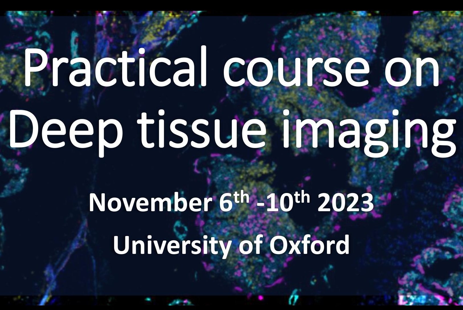
Practical course on Deep tissue imaging, Oxford, UK
Turku Cell Imaging and Cytometry Core Facility has teamed up with the Oxford-Zeiss Centre of Excellence at the Kennedy Institute of Rheumatology at the University of Oxford to organize a joint “Practical Course on Deep Tissue Imaging”. This course will take place in Oxford, UK on November 6-10. Course Description:Deep imaging (>50 μm) in biomedical samples such as thick tissue sections, spheroids, and organoids with fluorescence microscopy has become a much more common quest by bio-medical researchers. Yet, such imaging is very challenging as a consequence of difficulties with deep penetrative staining, and with optical limitations including light penetration, light scattering, and refractive index mismatch. The aim of this course is to collectively explore the use of a range of state-of-the-art fluorescence microscopes that are available within the Oxford – ZEISS Centre of Excellence at the University of Oxford, in order to gain a better understanding of the advantages and…
ISIDORE PROJECT “AUTOMATIC PIPELINE FOR BRAIN AUTORADIOGRAPHY IMAGE ANALYSIS”
Turku BioImaging, in collaboration with Zuzana Čočková (Charles University, Prague) and Francisco Lopez-Picón (Turku PET Centre) developed the Mouse Brain Alignment Tool (MBAT), a Python-based software for processing autoradiographic (ARG) images of mouse brain tissue sections and analyzing them using topographical data from the Allen Institute’s Mouse Brain Atlas. The ARG images of mouse brain were from a study that investigates brain damage caused by COVID-19. This project was funded by the ISIDORe project of Euro-BioImaging through the Finnish Advanced Microscopy Node. In June 2023, Zuzana visited Turku BioImaging (TBI) and developed the software together with Junel Solis, one of TBI’s image data analysts. The image is taken by Joanna Pylvänäinen. “The developed pipeline for analysis of ARG images will increase reliability and reproducibility as well as enable end-users to focus on image analysis without technical interruptions”, explains Junel. The source code repository will soon be made public and open source. The project results were…

Turku Imaging Day 2023, Turku, Finland
Turku BioImaging together with the Field of View Research Program organizes the 1st edition of Turku Imaging Day, which will take place on Tuesday 3rd of October, 2023! Turku Imaging Day brings together researchers interested in imaging and aims to strengthen and grow the imaging community in Turku. The main pillars of the Turku Imaging Day are: 1) Excellent, easy-to-follow, scientific talks all about imaging; 2) Networking and making new connections; 3) Evening Get-Together to turn your new connections to new friendships. There are different options to participate in the event: I) Flash Talk Competition: Are you an efficient and concise speaker? Introduce your research in two minutes using slides that only contain images; no text allowed. II) Poster Competition: You don’t fancy being on the stage? Present your research in a more relaxed setting during the lunch break. You can make brand new posters or recycle already used ones as long as they relate to imaging. III) Matchmaking Session: Do you miss a…

CytoData symposium 2023, Helsinki, Finland
CytoData symposium and hackathon brings together researchers interested in image-based profiling of biological phenotypes. The symposium including workshops, presentations and a poster session is scheduled for the first two days of the event (Oct 23-24) followed by a hackathon for developers during the last two days of the event (Oct 25-26). The themes of the symposium are Single-cell profiling and Spatial profiling. CytoData 2023 is the annual event of the CytoData Society hosted by Institute for Molecular Medicine Finland (FIMM), University of Helsnki and University of Turku. Symposium Dates & Venue: October 23-24, 2023. University of Helsinki City Center Campus, Main Building Hackaton Dates & Venue: October 25-26, 2023. University of Helsinki Meilahti Campus in the Biomedicum complex. Confirmed speakers Anne Carpenter, Broad Institute Berend Snijder, ETH Zurich Leeat Keren, Weizmann Institute of Science Loïc Royer, Chan Zuckerberg Biohub Wei Ouyang, KTH Royal Institute of Technology Symposium Chairs Lassi Paavolainen, Institute for Molecular Medicine Finland (FIMM), University of…